What's New?
The 1999-2000 Beckman Scholars: Sarah Brienne McCarthy (Faulkner), JD
| Faculty Mentor: Professor Andrew D. Ellington Length of term: Summer 99, Spring 00, Summer 00; Fall 99 was spent in a Study Abroad program in Madrid, Spain. Honors & Awards: Dean's Honored Graduate (Spring 01); University Honors (Spring 99); Junior Fellows Program (2000 – 2001); Ralph Nelson Endowed Presidential Scholarship (2000 – 2001); James S. Mahon Memorial Scholarship (2000 – 2001); University of Texas Distinguished Scholar (1999, 2000); University Co-Op Award for Superior Scholarly or Creative Achievement (2001). Publications: Chu, T.C. ; Marks, J.W., III ; Lavery, L.A. ; Faulkner, S.B. ; Rosenblum, M.G. ; Ellington, A.D. ; Levy, M. "Aptamer:Toxin Conjugates that Specifically Target Prostate Tumor Cells" Cancer Research 66, no. 12 (2006): 5989-5992. Faulkner, S.B; Syrett H.A.; Davidson E.A.; Ellington, A.D. In-vitro evolution through PCR-mediated mutagenesis and recombination. In PCR Technology: Current Innovations, 2nd Edition; Weissensteiner, T., Griffin, H.G., Griffin, A.; CRC Press LLC: Washington D.C., 2004; 371-380.; Two legal publications. Where is she now? Graduated from UT Austin with a Bachelor of Arts in Biochemistry with Highest Honors, May 2001, and a Master of Arts in Biochemistry in December 2004, and a JD from Yale in 2008. Sarah clerked for the US Federal courts, was an Intellectual Property lawyer in San Francisco, moved to British Columbia, and is now back in California. How can I contact her?sarahbrienne at gmail dot com |
 |
Beckman research project in the Ellington group:
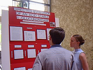
Directed evolution of an acid stable beta-glucuronidase
The protein beta-glucuronidase (GUS) is a found both in bacteria and in vertebrates. The vertebrate GUS homologue is most active under acidic conditions, demonstrating that certain mutations, alone or in combination, altered the pH profile of GUS during the course of evolution. The amino acid sequences of the E. coli and human GUS enzymes are only 50% identical, so the vast majority of differences are likely to be neutral with respect to this mechanism; however, it would be difficult to identify these mutations by a comparative structural approach. We therefore sought to identify mutations that may have played a role in shifting the pH optimum of the beta-glucuronidase protein. My objective was to evolve a bacterial protein that demonstrates a response to substrate at a lower pH, ideally a pH where the vertebrate version is shown to be stable, and to identify sequence substitutions that decrease the pH optimum of the E. coli GUS enzyme.
This was accomplished through rounds of random mutagenesis of the gusA gene, expression of the protein in E. coli, and a selection for activity in low pH buffer by a nitrocellulose filter-lift. The genetic sequence is first altered by PCR (polymerase chain reactions) to produce mutants. PCR is a process by which DNA can be actively produced by combining a template, nucleotides, and a polymerase in a buffered reaction mixture. To produce mutations in the sequence of the created DNA, the polymerase that places and links the dNTPs (nucleotides) is caused to be error-prone. The mutant genes are transformed into cells where the protein could be expressed. The colonies are then lysed by an exposure to chloroform, which allows endogenous lysozyme to digest the cell wall more easily. Colony remnants are screened to identify those containing GUS variants that are active in low pH. Through subsequent rounds of sequence alteration and screening, the amino acid substitutions that affect pH optima were revealed. The resulting mutant proteins were then purified, characterized, and sequenced.
After three rounds of mutagenesis and selection in decreasing pH, two variants were selected for comparison with the wild type. The round three variant, which also contains the mutations acquired in rounds one and two, was found to differ from the wild type by five amino acids substitutions: N181D, A208T, D508G, V355D, and V473A. The other variant, which was selected in an alternate round one screen at higher pH, contained one mutation, D508G, which is also contained in the round three variant. Two of the mutations, A208T and N181D, occur at the surface of the enzyme where they add positive charges. This phenomenon has been shown to increase enzyme activity by attracting substrate to the protein. The other three mutations, including D508G, are found within 12 angstroms of the active site and are probably involved in changing the pKa of amino acids essential for catalysis.
Steady-state kinetics was used to gain a quantitative comparison of the enzymes by comparing the reaction rates of the wild-type beta-glucuronidase protein versus the mutant beta-glucuronidase protein over a range of pH values. Enzyme activity was measured in saturating concentrations of pNP-glucuronide that absorbs at OD405 when the glycosidic bond is hydrolyzed by the GUS protein. Results were then compared for the wild type, round three variant, and the D508G mutant over a pH range of 7 to 3.5. The wild type exhibited a pH optimum of ~ 6.5, a sharp decline in activity from pH 5, and no detectable activity at pH 3.5. The round three variant showed little activity at pH 7 through 6, but did exhibit a sharp increase to reach an optimum at pH 4.0. The round three variant then experienced a sharp decline in activity to pH 3.5 where, like the wild type, detectable activity fell to zero. In contrast, the single mutant D508G, an amino acid change that is contained in the round three sequence, exhibited a very different pH profile. The D508G variant has a pH optima of 5, slightly more basic than the round three variant, but generally exhibits higher activity overall. Both variants did, however, show less activity than the wild-type for all pHs, except for pH 4.0 where both enzymes exceed the wild type by more than a factor of two. It would follow that with additional rounds of evolution, variant enzyme activity could be evolved to further exceed that of the wild type.
As a point of comparison between the wild type and our mutants, steady-state kinetics was also used to evaluate a vertebrate, bovine homologue. The pH optimum of the vertebrate enzyme was found to fall between 5.5 and 5.0, similar to our single mutant D508G. The activity, however, was found to be significantly less than that of the evolved variants. This result tends to contradict the belief that when evolution occurs, it is due primarily to the synergistic interaction of multiple amino acid substitutions.
I am preparing a first-author manuscript reporting on these results, which we intend to submit to the journal Protein Engineering within the next few months.

Created and maintained by Ruth Shear. Comments to author at DrRuth@mail.utexas.edu
Created Mon Mar 22nd 1999. Last modified Mon, Mar 16, 2015.