What's New?
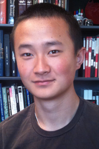
The 2011-2012 Beckman Scholars: Yafei Ouyang
| Faculty Mentor: Professor Christine E. Schmidt Length of term: Summer 11, Fall 11, Spring 12, Summer 12 Honors & Awards:University Honors (Fall 09, Spring 10, Fall 10, Spring 11, Fall 11); National Merit Finalist (2008);Chevron REACH scholarship (2008); Ernest J. Cockrell School of Engineering Scholarship (2008); University of Texas Co-op Society Undergraduate Research Fellowship (2010). Publications: Where is he now? Graduated with Bachelor of Science (Biomedical Engineering), May 2013. Yafei is currently at medical school at the Baylor College of Medicine. How can I contact him? yafei at utexas.edu |
 |
Beckman research project in the Schmidt group
Characterization of decellularized lung tissue using Vascular Corrosion Casting
I started the vascular corrosion casting project a little while before receiving the Beckman scholarship and throughout the extent of the scholarship mainly focused on this work. The following abstract was submitted to the World Conference on Regenerative Medicine in Leipzig for the oral presentation I gave detailing this project:
Researchers have used decellularization techniques as a direct way to try to mimic the native tissue architecture. Among the different methods, chemical decellularization techniques have been most often used due to ease in variation and ability to control the process. Thus different labs have developed unique chemical decellularization protocols for a host of different tissues.
In our experiment, we explored the effects of various decellularization techniques on lung tissue paying special attention to the preservation of vascular architecture. To do so, we applied three different decellularization techniques to murine lung tissue: the diffusive Hudson Optimized Acellularization (OA) process, the perfusion based Taylor Protocol (PD), and a hybrid perfusion Optimized Acellularization (POA) process. After decellularization, the tissues were analyzed using Vascular Corrosion Casting (VCC) to create polymer representations of the lumen space and vascular architecture. VCCs were imaged using a Scanning Electron Microscope (SEM). SEM images were analyzed with VCCs of fresh tissue as the negative control.
The control VCCs displayed lattice-like capillary networks and extensive larger vessel branching. Extravasations, formations resulting from the casting polymer leaking from hemorrhagic structures, were present but rare. The OA process resulted in VCCs that were the most similar in morphology to the fresh VCCs. Although there was not a complete lattice-like capillary structure, pseudo-capillary networks and extensive network branching were evident. The PD protocol resulted in casts in which irregular extravasations dominated and no signs of capillary structures were present. Larger vessels were retained though some showed damage and discontinuity. The hybrid POA process resulted in VCCs that showed a level of preservation between the OA and PD processes. Although no capillary structures were preserved, larger vessel branching networks were present and regular. Extravasations were common but uniform and limited to small spheroidal masses.
From these results we can conclude that the different decellularization methods are not equal. Though all these methods successfully remove cellular material, certain methods are far more damaging to the tissue than others. The PD protocol proved to be the harshest method whereas the OA process was the most gentle. Indeed, the OA process preserved most of the vascular network structure of the tissue. Thus although other processes were faster or more efficient, the slow OA process resulted in the best preserved tissue.
Since the conclusion of the vascular corrosion casting project, I have refocused my work on the larger project that my graduate student is pursuing. One of the largest challenges to developing tissue scaffolds for larger injury sites is the problem of tissue necrosis. The tissue often dies from the inside out as the cells outpace the growth of the vasculature, their only source of nutrients and waste removal. As such the goal of the project is to combat this problem by developing a pre-vascularized tissue scaffold that has an in vitro grown vascular network in place. To this end, murine lung tissue, a highly vascularized tissue, is used as the basis of the decellularized protein scaffold. Into this scaffold, endothelial progenitor cells are seeded and cultured under perfusion flow conditions to instigate formation of a patent vascular network.
Currently, we are in the process of perfusion culturing the cells in the scaffold. After the Vascular Corrosion Casting work, we decided to stick with the diffusion/optimized acellularization approach for creating the scaffolds. We then used photoacoustic imaging to investigate optimal cell seeding densities. This imaging technique allowed us to track the perfusion of cells that were marked with gold nanoparticles. With the cell seeding densities optimized, we are now focusing on developing a sterile and contained perfusion culture setup to culture the vascular network in vitro.
Created and maintained by Ruth Shear. Comments to author at DrRuth@mail.utexas.edu
Created Wed Jun 6th 2007. Last modified Mon, Mar 10, 2014.